Tendon Diagram / EPOS™ - C-1543 / The two peroneal tendons in the foot run side by side behind the outer ankle bone.. Attaches the calf muscles to the calcaneus, most important muscles for running, jumping, walking etc. This important tendon in the back of the calf and ankle connects the plantaris, gastrocnemius, and soleus muscles to. Learn about these muscles, their origin and insertion points, and their functional anatomy. Top (dorsal) view of foot & ankle number 1 and 2: Muscle anatomy gluteus 12 photos of the muscle anatomy gluteus gluteus muscle anatomy ct, gluteus muscle anatomy mri, human muscle anatomy gluteus maximus, muscle anatomy gluteus, muscle anatomy of gluteal, human muscles, gluteus muscle anatomy ct, gluteus muscle anatomy mri, human muscle anatomy gluteus maximus.
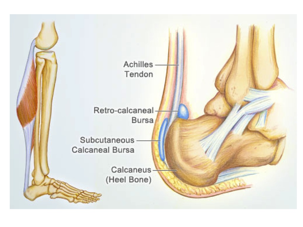
The achilles tendon is a tough band of fibrous tissue that connects the calf muscles to the heel bone (calcaneus). The two peroneal tendons in the foot run side by side behind the outer ankle bone. 2 ligaments (trapezoid& conoid ligaments) attach the clavicle coracoid process of scapula these tiny ligaments (w/ acominoclavicular joint) keep scapula attached to clavicle. Top (dorsal) view of foot & ankle number 1 and 2: Again, our knowledge of how mechanical stimulus mediates ligament and tendon structure is more empirical and less.
EPOS™ - C-1543 from epos.myesr.org It attaches to the wrist bone, the pisiform, and as well as the 5th hand bone. This important tendon in the back of the calf and ankle connects the plantaris, gastrocnemius, and soleus muscles to. Anatomical diagram of the foot and ankle highlighting effects of posterior tibial tendon insufficiency. Tendon, tissue that attaches a muscle to other body parts, usually bones. Arguably, the most important tendon is the achilles tendon, which allows the calf muscles to move the ankle joint. The ecu tendon works along with the ecrl and ecrb to straighten the wrist. Foot anatomy diagram, foot joint diagram, foot sprain diagram, foot tendons and ligaments pain, leg tendon diagram, peroneal tendonitis, foot, foot anatomy diagram, foot joint diagram, foot sprain diagram, foot tendons and ligaments pain, leg tendon diagram, peroneal tendonitis. The pubis, ischium, and ilium together constitute the pelvis while the thigh bone is the femur.
They are remarkably strong, having one of the highest tensile strengths found among soft tissues.
The achilles tendon is a tough band of fibrous tissue that connects the calf muscles to the heel bone (calcaneus). Observe the leg muscle diagram posted above and notice that there are many parts in the muscles.the largest muscle masses in the leg are present in the thigh and the calf. The ecu tendon works along with the ecrl and ecrb to straighten the wrist. The bones of the hip include the femur, the ilium, the ischium, and the pubis. The rotator cuff is a group of four muscles and tendons that surround the glenohumeral joint. Tendons attach muscles to bones. Muscle anatomy gluteus 12 photos of the muscle anatomy gluteus gluteus muscle anatomy ct, gluteus muscle anatomy mri, human muscle anatomy gluteus maximus, muscle anatomy gluteus, muscle anatomy of gluteal, human muscles, gluteus muscle anatomy ct, gluteus muscle anatomy mri, human muscle anatomy gluteus maximus. It attaches to the wrist bone, the pisiform, and as well as the 5th hand bone. Tendons are similar to ligaments; Again, our knowledge of how mechanical stimulus mediates ligament and tendon structure is more empirical and less. Ultrasound can often diagnose an achilles tendon rupture. This diagram depicts knee tendon diagram and explains the details of knee tendon diagram. Each of these muscles is a discrete organ constructed of skeletal muscle tissue.
Muscles of the shoulder : Flexor tendon lacerations are classified into five zones 2, 15, 16. Human hand tendon diagram (page 1) hand tendons diagram muscle blank drawing these pictures of this page are about:human hand tendon diagram this small muscle is located at the top of the shoulder and helps raise the arm away from the body. The achilles tendon is a tough band of fibrous tissue that connects the calf muscles to the heel bone (calcaneus). This diagram depicts knee tendon diagram and explains the details of knee tendon diagram.
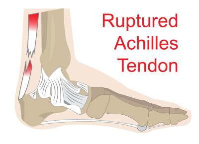
Achilles Tendon Ruptures | Issaquah Foot & Ankle Specialists from images.fosterwebmarketing.com The achilles tendon enables us to walk, without it we would not be able to raise our heels of the ground. This important tendon in the back of the calf and ankle connects the plantaris, gastrocnemius, and soleus muscles to. Muscles of the shoulder : One peroneal tendon attaches to the outer part of the midfoot, while the other tendon runs under the foot and attaches near the inside of the arch. Flexor tendon lacerations are classified into five zones 2, 15, 16. Diagram showing the tendons and ligaments of the ankle and. The achilles tendon is the largest. Fall on one point of shoulder and can rupture these ligaments with dislocation of ac joint.
The coracobrachialis muscle lies deep to the biceps brachii in the arm.
Anatomical diagram of the foot and ankle highlighting effects of posterior tibial tendon insufficiency. Tendons attach muscles to bones. Ligaments and tendons are adapted in response to changes in mechanical stiffness. 17 photos of the diagram of shoulder muscles and tendons. The tendon runs down the back of your lower leg from the back of the knee to the heel. Tendons are the connection between bones and muscles. The coracobrachialis muscle lies deep to the biceps brachii in the arm. The rotator cuff is a group of four muscles and tendons that surround the glenohumeral joint. Ligaments and tendons are fibrous connective tissues made up of densely packed collagen fibers. Ultrasound can often diagnose an achilles tendon rupture. Muscle anatomy gluteus 12 photos of the muscle anatomy gluteus gluteus muscle anatomy ct, gluteus muscle anatomy mri, human muscle anatomy gluteus maximus, muscle anatomy gluteus, muscle anatomy of gluteal, human muscles, gluteus muscle anatomy ct, gluteus muscle anatomy mri, human muscle anatomy gluteus maximus. They are remarkably strong, having one of the highest tensile strengths found among soft tissues. One peroneal tendon attaches to the outer part of the midfoot, while the other tendon runs under the foot and attaches near the inside of the arch.
The rotator cuff is a group of four muscles and tendons that surround the glenohumeral joint. The biceps muscle has two tendon attachments. 17 photos of the diagram of shoulder muscles and tendons. This important tendon in the back of the calf and ankle connects the plantaris, gastrocnemius, and soleus muscles to. Ligaments and tendons are adapted in response to changes in mechanical stiffness.
Unit 2 Anatomy Diagrams - Biology Biol&241 with Dr. Vega ... from s3.amazonaws.com Diagram showing the tendons and ligaments of the ankle and. The two peroneal tendons in the foot run side by side behind the outer ankle bone. Top (dorsal) view of foot & ankle number 1 and 2: The hip itself is a ball and socket joint, much like the shoulder.the structures necessary to create this joint are the socket, the joint capsule, muscle, ligaments, and the neck. The ecu tendon works along with the ecrl and ecrb to straighten the wrist. This tendon connects the patella (kneecap) to the tibia. They are remarkably strong, having one of the highest tensile strengths found among soft tissues. Also allows the action of raising up onto toes.
Foot anatomy diagram, foot joint diagram, foot sprain diagram, foot tendons and ligaments pain, leg tendon diagram.
The hip itself is a ball and socket joint, much like the shoulder.the structures necessary to create this joint are the socket, the joint capsule, muscle, ligaments, and the neck. Learn about the anatomy and physiology of tendons. Bones, cartilage, ligaments, and tendons. Foot anatomy diagram, foot joint diagram, foot sprain diagram, foot tendons and ligaments pain, leg tendon diagram, peroneal tendonitis, foot, foot anatomy diagram, foot joint diagram, foot sprain diagram, foot tendons and ligaments pain, leg tendon diagram, peroneal tendonitis. The largest of these shoulder muscles is the. Superficial posterior muscles of the forearm posterior compartment muscles of the forearm. The rotator cuff is a group of four muscles and tendons that surround the glenohumeral joint. Related posts of muscles and tendons of the leg muscle anatomy gluteus. When the muscles tighten (contract) arguably, the most important tendon is the achilles tendon, which allows the calf muscles to move. The muscles that make up the quadriceps are the strongest and leanest of all muscles in the body. The achilles tendon is the largest. You can see a diagram of the achilles tendon below. A tendon is a band of tissue that connects a muscle to a bone.